Intro
Week 6 of pregnancy is often the moment when an early pregnancy begins to feel more tangible. You may be navigating nausea, breast tenderness, fatigue, mood changes, or the anxiety of waiting for a first scan. At the same time, inside the uterus, the embryo is undergoing rapid, highly coordinated development, especially in the heart, neural tube, early limb buds, and organ primordia.
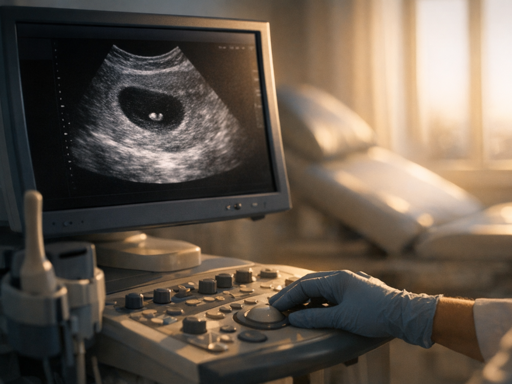
Medically, week 6 is also a nuanced time. Ultrasound may show a gestational sac, yolk sac, fetal pole, and sometimes cardiac activity, but the exact findings depend strongly on dating accuracy, ovulation timing, equipment, scan route, and embryo size. A scan that is “too early” can be emotionally unsettling even when the pregnancy is developing normally, so results should always be interpreted with a qualified clinician.
Highlights
By week 6, the embryonic heart is no longer just a simple tube; it is actively looping and beginning to form more complex early chambers and outflow structures.
A heartbeat may be visible on transvaginal ultrasound around 6 to 7 weeks, but absence of visible cardiac activity at a single early scan does not always mean pregnancy loss.
Organ formation is accelerating: the neural tube, early brain regions, limb buds, facial structures, gut, liver primordium, and early circulation are all developing.
Symptoms can be intense or surprisingly mild at week 6; symptom severity alone is not a reliable measure of pregnancy viability.
Prompt medical attention is important for heavy bleeding, severe one-sided pain, shoulder-tip pain, fainting, fever, or worsening symptoms that feel unsafe.
How pregnancy dating works in week 6
Pregnancy are counted from the first day of the , not from conception. In a typical 28-day cycle, week 6 of pregnancy corresponds to about four after fertilization. However, many people ovulate earlier or later than day 14, and implantation timing also varies. This is why a pregnancy labeled “6 weeks” by menstrual dates may look closer to 5 weeks or 6 weeks 5 days on ]].
This distinction matters because nic structures change quickly from day to day. A gestational sac may be visible before a yolk sac, a yolk sac before a pole, and a pole before cl detectable cardiac activity. A few days can substantially change what is seen. When findings are uncertain, clinicians often recommend repeat and serial blood tests rather than making conclusions from one image.
Heart development: from primitive tube to early rhythm
The nic heart is one of the first functional organs. By the sixth week, it has already begun beating and circulating blood through the embryo’s early vascular network. Developmentally, the heart begins as a primitive tube that folds, loops, and partitions over time. During week 6, this tube is transforming into a more complex structure, with early inflow and outflow regions and developing aortic arch structures.
Cardiac looping is especially important. It helps establish the future left-right orientation of the heart and lays the foundation for later chamber formation. Septation, valve development, and maturation of the conduction system will continue over the coming weeks, but week 6 is a critical period for the basic architecture of the cardiovascular system.
On ultrasound, cardiac activity may appear as a tiny flicker within the fetal pole. At this stage, it is usually too early to assess detailed heart anatomy. The clinical question is typically whether cardiac activity is present and whether the embryo’s size and heart rate are appropriate for the estimated gestational age. A peer-reviewed study on embryonic heart rate in the early showed that normal ranges vary by gestational age and that interpretation around week 6 requires careful dating rather than a single universal cutoff.
What heart rate may be expected around 6 weeks
When cardiac activity is detected at 6 to 7 weeks, a commonly cited early range is approximately 90 to 110 beats per minute, although values rise as the first trimester progresses. Early heart rate is dynamic; it tends to increase over the next several weeks before later stabilizing. A heart rate that seems slow or uncertain very early may need follow-up rather than immediate interpretation in isolation.
Several factors can affect what is measured:
- Gestational age accuracy: Being off by even three to five days can change expectations.
- Crown-rump length: Heart-rate interpretation is more meaningful when paired with embryo size.
- Scan type: Transvaginal usually detects early structures sooner than abdominal .
- Image quality: Uterine position, body habitus, equipment, and operator experience can all influence visibility.
- Measurement variability: Very small structures are harder to measure precisely.
If your report mentions a heart rate that is lower than expected, higher than expected, or not yet measurable, it is understandable to feel worried. The safest next step is to review the result with your obstetric clinician, who can place it in context and decide whether repeat imaging is appropriate.
Organ formation and embryonic growth in week 6
Week 6 is part of the nic period, when organogenesis is highly active. The is still very small, but its al workload is extraordinary. The neural tube, which will give rise to the brain and spinal cord, is closing and differentiating. Early brain vesicles are forming, and the head region appears relatively large because brain is so prominent.
Other major changes include:
- Limb buds: Small upper and lower limb buds begin to emerge, laying the groundwork for arms and legs.
- Facial ]]: Early structures that will contribute to the eyes, ears, jaw, and face are forming.
- Digestive tract: The primitive gut tube is developing and will later differentiate into the grointestinal tract.
- Liver and blood formation: The early liver region becomes important in blood-cell production during embryonic.
- Circulation: Blood vessels are expanding as the heart supports early circulation.
- Placental support: The placenta and chorionic villi continue developing, although the placenta is not yet fully mature.
Because organ formation is so active, week 6 is also a time when general prenatal health matters. If you have not already done so, ask your healthcare professional about prenatal vitamins, folic acid intake, medication safety, chronic condition management, and avoidance of alcohol, nicotine, and non-prescribed substances. Do not stop prescribed medication abruptly without advice; for many conditions, untreated illness can also carry risk.
Ultrasound expectations at week 6
A 6-week is usually performed transvaginally if the goal is to evaluate early viability, dating, , pain, or prior pregnancy loss. Abdominal ultrasound may be used in some settings, but it is generally less sensitive this early. A typical scan may look for the location of the pregnancy, number of gestational sacs, yolk sac, fetal pole, crown-rump length, and cardiac activity if visible.
Possible findings include:
- Gestational sac: A fluid-filled structure within the uterus, often visible before the embryo is seen.
- Yolk sac: An early support structure that helps confirm an intrauterine pregnancy when seen in the gestational sac.
- Fetal pole: The early embryo, measured by crown-rump length.
- Cardiac activity: A flickering motion that may be detected around 6 to 7 weeks, especially with transvaginal imaging.
- Dating estimate: Ultrasound dating may differ from menstrual dating, particularly with irregular cycles.
It is also possible that the scan is inconclusive. For example, a gestational sac and yolk sac may be seen without a fetal pole, or a fetal pole may be seen without definite cardiac activity. Depending on measurements and symptoms, clinicians may repeat the ultrasound in 7 to 14 days. That waiting period can be emotionally difficult, but it helps avoid misclassifying a pregnancy that is simply earlier than expected.
Common symptoms in week 6
Hormonal changes, especially rising human chorionic gonadotropin and progesterone, can make week 6 physically demanding. Nausea may intensify, sometimes with food aversions, increased saliva, bloating, constipation, breast tenderness, frequent urination, and profound fatigue. Mild cramping can occur as the changes and the corpus luteum supports early pregnancy.
Symptom patterns vary widely. Some feel very pregnant; others have minimal symptoms. A sudden change in symptoms can be alarming, but symptoms alone cannot confirm whether a pregnancy is progressing normally. Conversely, severe nausea and vomiting may require medical care, particularly if you cannot keep fluids down, are losing weight, or have signs of dehydration.
Emotional symptoms are also real. Early pregnancy after infertility, miscarriage, ectopic pregnancy, or medical complications can bring intense vigilance. If you find yourself repeatedly checking symptoms or feeling overwhelmed while waiting for results, it may help to ask your clinic what changes truly require a call and when follow-up is planned.
How to support health during this stage
Supportive care at week 6 focuses on reducing avoidable risks and making sure medical issues are managed. General steps include taking a prenatal vitamin as advised, ensuring adequate folic acid intake, staying hydrated, eating small tolerable meals if nauseated, and getting rest when possible. Gentle is usually acceptable for many pregnancies, but individual recommendations may differ for bleeding, pain, fertility treatment pregnancies, or specific medical conditions.
Contact healthcare professional about medication review, occupational exposures, travel concerns, infectious disease risks, and any chronic conditions such as diabetes, thyroid disease, epilepsy, hypertension, autoimmune disease, or mental health conditions. Early pregnancy is not the time to manage these questions alone; small adjustments can be important, and individualized guidance is safer than generic advice.
If you have an upcoming , consider asking what the clinic expects to see based on ]] dates, whether the scan will be transvaginal, what findings would trigger repeat imaging, and how results will be communicated. Clear expectations can reduce uncertainty and help you feel more prepared.
When to seek urgent medical advice
- Heavy bleeding, passing large clots, or soaking pads rapidly should be assessed promptly.
- Severe one-sided pelvic pain, shoulder-tip pain, dizziness, or fainting can be concerning and needs urgent evaluation.
- Fever, chills, or severe abdominal pain should be discussed with a healthcare professional immediately.
- Persistent vomiting with inability to keep fluids down may require treatment for dehydration.
- If you have a history of ectopic pregnancy or are at increased risk, report pain or bleeding early.
Tools & Assistance
- Schedule or confirm an early prenatal appointment with an obstetric clinician or midwife.
- Keep a list of medications, supplements, allergies, and medical conditions for review.
- Use a symptom and bleeding log if your clinician has asked you to monitor changes.
- Ask your clinic whether transvaginal ultrasound or repeat imaging may be needed.
- Seek urgent care or emergency services for severe pain, fainting, or heavy bleeding.
FAQ
Should a heartbeat always be seen at 6 weeks?
Not always. Cardiac activity may be seen around 6 to 7 weeks, especially with transvaginal ultrasound, but late ovulation or early dating can make a scan inconclusive.
What is a normal heart rate at 6 weeks?
A commonly cited range around 6 to 7 weeks is about 90 to 110 beats per minute, but interpretation depends on exact gestational age, crown-rump length, and clinical context.
Can organ problems be detected on a 6-week ultrasound?
Usually not in detail. Week 6 ultrasound focuses on pregnancy location, dating, early structures, and cardiac activity rather than detailed anatomy.
Is mild cramping normal at week 6?
Mild, intermittent cramping can occur in early pregnancy. Severe, persistent, or one-sided pain, especially with bleeding or dizziness, should be evaluated urgently.
What if my symptoms suddenly decrease?
Symptoms can fluctuate and are not a reliable diagnostic tool. If you are worried, especially with bleeding or pain, contact your healthcare professional.
Sources
- PubMed — Embryonic heart rate in the early first trimester
- American Pregnancy Association — Early Fetal Development: Trimester 1 Milestones
- Charlotte Lozier Institute — Week 6
Disclaimer
This article is for informational purposes only and does not replace professional medical advice, diagnosis, or treatment. Always consult a qualified healthcare professional about pregnancy symptoms, ultrasound findings, or personal medical decisions.