Intro
Pregnancy is usually dated from the first day of the last menstrual period and spans about 40 weeks, although individual pregnancies vary. Month-by-month milestones can make this timeline feel more understandable: the earliest weeks focus on implantation and organ formation, the middle months bring rapid growth and sensory development, and the final months prepare the fetus for life outside the uterus.
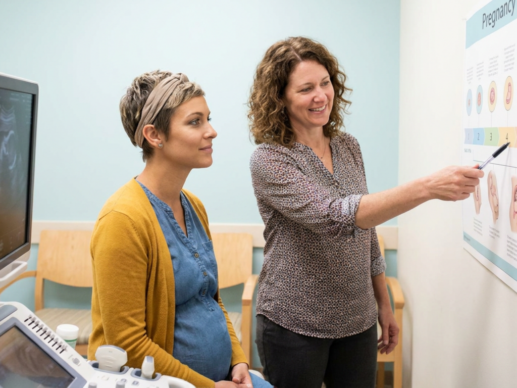
This guide summarizes typical fetal and maternal milestones by month. It is meant to support informed conversations with your obstetrician, midwife, family physician, or maternal-fetal medicine specialist, not to replace individualized prenatal care.
Highlights
The first trimester is a period of intense embryologic development, including neural tube closure, early cardiac activity, limb buds, facial structures, and the transition from embryo to fetus.
The second trimester often brings more visible pregnancy changes, fetal movement, detailed anatomy assessment, skeletal growth, and increasing sensory function.
The third trimester is dominated by fetal weight gain, brain and lung maturation, positioning for birth, and close monitoring of maternal and fetal well-being.
Milestone timing is approximate; ovulation date, ultrasound dating, fetal position, placental location, and individual variation can all affect what is seen or felt at a given visit.
How pregnancy months relate to weeks and trimesters
Pregnancy is commonly divided into three trimesters: weeks 1 to 12, weeks 13 to 27, and weeks 28 to birth. Month-by-month descriptions are approximate because calendar months do not align perfectly with gestational weeks. Clinically, your care team will usually communicate in weeks and days, such as 20 weeks 3 days.
A due date is an estimate, not a deadline. Many pregnancies continue safely beyond 40 weeks with appropriate monitoring, while some babies are born earlier for spontaneous or medically indicated reasons. Ultrasound dating in early pregnancy can help refine gestational age, especially when menstrual dates are uncertain.
Month 1: fertilization, implantation, and the earliest signals
Month 1 generally includes weeks 1 through 4. In standard obstetric dating, the first two weeks occur before conception, beginning with the last menstrual period. Fertilization typically occurs around ovulation, followed by cell division and travel through the fallopian tube. The developing blastocyst implants into the uterine lining, and trophoblast cells begin producing human chorionic gonadotropin, or hCG, the hormone detected by most pregnancy tests.
Development is microscopic but biologically profound. The early embryonic disc begins organizing into layers that will give rise to major tissues and organs. Some people notice breast tenderness, fatigue, mild cramping, mood shifts, nausea, or a missed period, while others feel little different. Light spotting can occur around implantation, but any heavy bleeding, severe pain, or shoulder pain warrants prompt medical evaluation.
Month 2: neural tube closure, early heartbeat, and limb formation
Month 2, approximately weeks 5 through 8, is one of the most developmentally sensitive periods. The neural tube, which becomes the brain and spinal cord, closes early in this window. The heart begins forming and may show early cardiac activity on ultrasound. Limb buds appear, the beginnings of eyes and ears develop, and facial structures start taking shape.
By the end of this month, major organ systems have begun forming, although they are far from mature. The embryo is still very small, but growth is rapid. Many pregnant people experience intensifying nausea, food aversions, fatigue, urinary frequency, and breast changes due to rising pregnancy hormones. If vomiting prevents hydration, if weight loss is significant, or if symptoms feel unmanageable, professional guidance is important.
Month 3: transition from embryo to fetus and early movement
Month 3 roughly covers weeks 9 through 12. Around this time, the developing baby is typically referred to as a fetus rather than an embryo. The head remains proportionally large, the face becomes more defined, fingers and toes are more distinct, and early bones and muscles continue developing. External genital structures begin differentiating, although ultrasound determination of fetal sex may still be unreliable this early.
The fetus may make small spontaneous movements, but these are usually too subtle to be felt. Kidneys begin producing urine, and reflex-like activity starts emerging. For the pregnant person, nausea may peak and then gradually improve near the end of the first trimester, though this varies. Prenatal care may include dating ultrasound, blood type and antibody screening, infectious disease screening, and discussion of genetic screening options.
Month 4: rapid growth, stronger skeleton, and a more visible pregnancy
Month 4, approximately weeks 13 through 16, begins the second trimester for most clinical frameworks. The risk profile changes as early organ formation is largely complete, though development and maturation continue. The fetus grows quickly, the neck becomes more defined, limbs lengthen, and the skeleton continues ossification, meaning cartilage is gradually replaced by bone.
Some people begin to feel better as nausea and fatigue ease, while others continue to have significant symptoms. A growing uterus may become more noticeable above the pelvis. Round ligament discomfort, constipation, nasal congestion, and skin changes can occur. If you have chronic conditions such as hypertension, diabetes, thyroid disease, autoimmune disease, or epilepsy, this is a key time to keep care coordinated with your pregnancy clinician and relevant specialists.
Month 5: anatomy scan, fetal movement, and sensory development
Month 5 usually includes weeks 17 through 20. Many people feel the first fetal movements, often called quickening, during this period. At first, movement may feel like fluttering, bubbles, or gentle taps. People who have been pregnant before may notice movement earlier, while an anterior placenta can make movement harder to perceive initially.
A major milestone is the mid-pregnancy anatomy ultrasound, commonly performed around 18 to 22 weeks. This scan evaluates fetal anatomy, growth, placental location, amniotic fluid volume, and sometimes cervical length depending on risk factors and local practice. The fetus is developing more coordinated movements, swallowing amniotic fluid, and continuing musculoskeletal growth. This halfway point can feel emotionally significant, especially after infertility, pregnancy loss, or medical complications.
Month 6: lung branching, hearing, and increasing viability discussions
Month 6, approximately weeks 21 through 24, brings continued growth of the lungs, blood vessels, and nervous system. The lungs are not mature, but airway branching and early structures needed for gas exchange are developing. The fetus may respond to sound and may develop patterns of activity and rest. Skin remains thin and translucent, with fat stores still limited.
Clinically, this period may include screening for gestational diabetes soon after, depending on local timing and individual risk. Some clinicians also assess anemia, blood pressure, and symptoms of preterm labor. The concept of viability may be discussed in high-risk situations, but outcomes at extremely early gestational ages depend on many factors and require individualized counseling from neonatal and obstetric specialists.
Month 7: third trimester begins and fetal activity becomes more patterned
Month 7 roughly spans weeks 25 through 28 and marks the transition into the third trimester. Brain development accelerates, eyelids can open, and the fetus continues accumulating fat. Lung maturation progresses, although more time in utero remains important. Movements often become stronger and more recognizable, including kicks, rolls, stretches, and hiccup-like rhythmic sensations.
Maternal changes may include heartburn, back or pelvic discomfort, leg cramps, sleep disruption, Braxton Hicks contractions, and shortness of breath as the uterus enlarges. This is also a common time for Rh immune globulin if the pregnant person is Rh-negative and clinically eligible, as well as additional blood tests or vaccines depending on medical guidance. Ask your care team when and how they want you to monitor fetal movement.
Month 8: weight gain, brain growth, and preparation for birth
Month 8, approximately weeks 29 through 34, is characterized by substantial fetal weight gain, increasing subcutaneous fat, and ongoing maturation of the brain and lungs. The fetus may settle into a head-down position, although position can still change. The amount of amniotic fluid, placental function, and fetal growth may be monitored more closely if there are risk factors such as hypertension, diabetes, fetal growth restriction, multiple pregnancy, or prior complications.
For the pregnant person, physical demands often intensify. Pelvic pressure, swelling of the feet or ankles, carpal tunnel symptoms, reflux, and fatigue are common, but sudden severe swelling, headache, visual symptoms, or right upper abdominal pain can suggest a serious condition such as preeclampsia and should be assessed urgently. Birth planning becomes more practical now: choosing support people, understanding labor signs, and discussing preferences for pain relief, feeding, and newborn care.
Month 9: final maturation, positioning, and readiness for labor
Month 9 generally includes weeks 35 through 40. The fetus continues gaining weight, the brain grows rapidly, and the lungs continue maturing. By 37 weeks, a pregnancy is generally considered early term; full term begins at 39 weeks. These distinctions matter because important maturation continues even late in pregnancy.
The fetus often moves lower into the pelvis, sometimes called lightening, although this may happen close to labor or not noticeably at all. Movements may feel different because space is tighter, but they should not significantly decrease. Prenatal visits typically focus on blood pressure, fetal heart rate, fetal position, growth concerns, symptoms, and planning for labor or induction if indicated. Group B streptococcus screening is commonly performed in late pregnancy according to local guidelines.
Milestones are reassuring, but individual variation is normal
It is natural to compare your pregnancy with charts, apps, and other people’s experiences. Still, milestone timing can vary. A fetal movement felt at 18 weeks versus 21 weeks may both be normal. A scan may need to be repeated because fetal position limits views. A fundal height measurement may differ slightly from expected and still not indicate a problem.
The most useful approach is to combine general knowledge with individualized care. Bring questions to prenatal visits, clarify which warning signs apply to your situation, and ask how your medical history affects monitoring. If something feels wrong, it is always appropriate to contact your healthcare team rather than waiting for the next scheduled appointment.
When to seek urgent medical advice
- Heavy vaginal bleeding, severe abdominal pain, fainting, or shoulder pain.
- Persistent vomiting with inability to keep fluids down or signs of dehydration.
- Severe headache, visual changes, sudden swelling, chest pain, or shortness of breath.
- Regular painful contractions, leaking fluid, or pelvic pressure before term.
- A noticeable decrease in fetal movement after movement patterns are established.
- Fever, painful urination, or symptoms that feel severe, sudden, or concerning.
Tools & Assistance
- Keep a written list of questions for each prenatal visit.
- Use a pregnancy calendar or app that tracks gestational weeks rather than only months.
- Attend recommended prenatal appointments, ultrasounds, and screening tests.
- Ask your clinician for individualized guidance on fetal movement monitoring.
- Know your maternity unit or emergency contact pathway before the third trimester.
FAQ
Which month is the most critical for fetal development?
The first trimester, especially months 2 and 3, is critical because major organs and the neural tube form early. However, growth and maturation remain important throughout pregnancy.
When should I feel the baby move?
Many people first feel movement between about 18 and 22 weeks, though timing varies. Contact your clinician if you are concerned or if established movement patterns decrease later in pregnancy.
Is the anatomy scan always done in month 5?
It is commonly scheduled around 18 to 22 weeks, which often falls in month 5. Timing may vary based on local practice, fetal position, pregnancy dating, and medical needs.
Does reaching 37 weeks mean the baby is fully mature?
At 37 weeks, pregnancy is considered early term, but important brain, lung, and weight gain milestones continue. Full term generally begins at 39 weeks.
What if my milestones do not match a month-by-month chart?
Small differences are common. Dating uncertainty, placental position, fetal position, and individual variation can affect symptoms and scan findings. Discuss specific concerns with your care team.
Sources
- Mayo Clinic — Fetal development: The first trimester
- Office on Women's Health — Stages of pregnancy
- Cleveland Clinic — Fetal Development: Week-by-Week Stages of Pregnancy
Disclaimer
This article is for informational purposes only and is not a substitute for professional medical advice, diagnosis, or treatment. Always consult a qualified healthcare professional about pregnancy symptoms, screening, medications, or urgent concerns.


Please log in to leave a comment.