
Intro
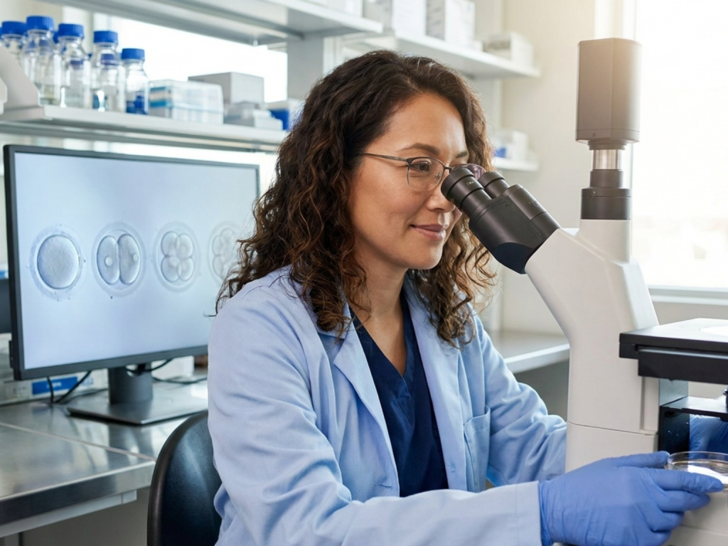
After fertilization, the fertilized egg does not simply sit unchanged; it enters a tightly coordinated developmental program. The single-cell zygote begins a series of rapid mitotic divisions called cleavage, which distribute the original cytoplasm into many smaller cells while preserving the embryo inside the zona pellucida.
For many people, this stage feels abstract because it happens so early, often before a pregnancy can be seen on ultrasound or confirmed by symptoms alone. Understanding the biology can make the early timeline feel less mysterious and can also clarify why implantation, pregnancy testing, and early loss are all linked to what happens in these first few days.
Highlights
The first divisions after fertilization are mitotic cleavage divisions, not ordinary growth divisions. They split one zygote into many smaller cells called blastomeres.
Early development includes a major cellular reset: the oocyte completes meiosis, then the embryo switches into the mitotic cycle that supports growth and organization.
Cell number rises quickly, but overall embryo size changes very little at first because the embryo is still enclosed by the zona pellucida.
Around the 8- to 16-cell stage, cells begin compaction, a structural reorganization that helps the embryo progress toward the morula and then the blastocyst.
Not every fertilized egg becomes a pregnancy; many early arrests are due to chromosomal or developmental issues that cannot be detected from symptoms alone.
What changes right after fertilization
Fertilization creates a zygote, but that is not the end of the story; it is the beginning of a major cellular transition. In the oocyte, the chromosomal program is built for meiosis, the special division that produces a mature egg. After sperm entry, the egg completes meiosis and then reorganizes itself for mitosis, the division pattern that generates identical daughter cells. This shift from meiosis to mitosis is one of the defining events of the earliest embryo.
At the same time, the embryo restores the machinery needed for division, including spindle organization and centrosome function. In humans, sperm-derived components help the newly formed embryo coordinate its first mitotic spindle. The maternal and paternal genetic material do not remain permanently separate; instead, they are brought into a common developmental program that prepares the zygote for the first cleavage divisions.
These changes are highly ordered, which is why this stage is often described as the embryo being biologically re-started. The cell is no longer just an egg or a sperm product; it is now a new organism beginning its own developmental sequence.
Cleavage: division without overall growth
The earliest mitotic divisions after fertilization are called cleavage. This term matters because cleavage is not the same as ordinary growth. The embryo divides again and again, but the total volume changes little at first. Instead of becoming larger with each division, the original cytoplasm is partitioned into smaller cells. That is why the cells are called blastomeres.
The zona pellucida, the protective outer shell surrounding the early embryo, helps explain this pattern. Because the embryo remains enclosed, the new cells have limited room to expand. As a result, cell number rises while individual cell size falls. This is a crucial design feature of early development: the embryo is creating cellular units and organizing them, not yet building bulk.
Medically, it is helpful to think of cleavage as a countdown of cellular reorganization. One cell becomes two, then four, then eight, and so on. The process is fast, but it is also coordinated, and it sets up the later steps that allow the embryo to implant and continue developing. The first zygote cleavage divisions are therefore a structural preparation for pregnancy, not a sign of pregnancy progression by themselves.
From two cells to morula
As cleavage continues, the embryo moves through recognizable stages. A one-cell zygote becomes a two-cell embryo, then a four-cell embryo, and then a larger cluster of small blastomeres. By the time the embryo reaches roughly the 8- to 16-cell range, it starts to compact. Compaction means the cells flatten against one another, strengthen their cell-to-cell adhesion, and begin to look less like isolated spheres and more like an integrated mass.
This is the stage often described as morula formation after fertilization. The morula is a solid ball of cells, and it represents an important shift in organization. The cells are no longer just dividing; they are beginning to differentiate in behavior and position. Some cells become more central, while others stay on the outside, and this spatial arrangement helps prepare the embryo for the next phase.
These changes are not visible to a person in everyday life, but they are biologically meaningful. If a fertilized egg reaches the morula stage, it has passed through several successful rounds of division and structural coordination. At the same time, early arrest can occur before or during this phase, often because of chromosomal errors that are common in human reproduction.
Morula to blastocyst: preparing for implantation
After the morula, the embryo reorganizes again and becomes a blastocyst. This step is important because the blastocyst is the form that can eventually implant in the uterus. The outer cells begin to specialize into trophoblast lineages, while an inner cell mass remains available for forming the embryo proper. In other words, the embryo is separating future support structures from future fetal tissues.
The blastocyst stage before implantation is also when the embryo starts to handle fluid differently, creating a cavity called the blastocoel. That hollow space marks a more advanced level of organization than the solid morula. Soon after, the embryo must hatch from the zona pellucida so it can interact directly with the endometrium.
It is reassuring to know that implantation does not happen immediately after fertilization. A sequence of successful divisions, compaction, and blastocyst formation must occur first. For that reason, early pregnancy is best understood as a progression rather than a single event. Fertilization starts the process, but implantation is the later milestone that establishes an ongoing pregnancy.
Why early cell division can vary
Although embryology textbooks often present the sequence in a neat order, real human development is more variable. The timing and appearance of cleavage divisions can differ from one embryo to another. Some embryos divide smoothly, while others show slower progression or uneven cleavage patterns. These differences do not automatically reveal whether a pregnancy will continue, and they cannot be assessed accurately from symptoms alone.
One reason for variation is that early development depends on precise chromosome segregation and cellular coordination. If chromosomal abnormalities are present, the embryo may stop dividing or may fail to progress beyond the earliest stages. This is one reason many conceptions do not reach implantation, even when fertilization has occurred. It is biologically common, and it is not something a person usually feels.
Because of this variability, clinicians interpret early pregnancy using the full clinical picture rather than a single sign. The biology of cleavage can explain why timing differs, but only a healthcare professional can evaluate whether a specific situation needs observation, testing, or follow-up.
Why this stage matters for pregnancy care
Understanding cell division after fertilization helps connect the invisible early days of pregnancy with later milestones such as implantation and pregnancy testing. Before hCG becomes detectable, the embryo must pass through cleavage, compaction, and blastocyst formation. That means there is a biologic gap between fertilization and the point at which a pregnancy test can turn positive.
This sequence also explains why the earliest stage of pregnancy is so vulnerable. A fertilized egg may be present, yet the embryo may not be able to continue dividing or implant. That is not a failure of effort; it is part of the natural biology of early reproduction. Many people find that framing helpful because it replaces self-blame with a clearer understanding of how delicate early embryonic development is.
If you are trying to conceive, waiting to test, or trying to interpret early symptoms, it can help to think in stages: fertilization, zygote cleavage divisions, morula formation after fertilization, blastocyst stage before implantation, and then implantation itself. That framework can make discussions with a clinician more precise and less stressful.
When to seek medical advice
- Severe one-sided pelvic pain, heavy bleeding, fainting, or shoulder pain need urgent medical assessment.
- If you have a history of recurrent pregnancy loss or fertility treatment, ask your clinician how early development is being monitored.
- A positive or negative pregnancy test does not by itself explain what happened at the cellular level.
- Do not assume that a symptom pattern proves normal development or early loss; evaluation matters.
Tools & Assistance
- Obstetrician-gynecologist consultation for early pregnancy questions
- Reproductive endocrinology clinic if you are being evaluated for fertility concerns
- Early pregnancy ultrasound when a clinician says it is appropriate
- Laboratory hCG testing through a healthcare service if recommended
- Patient education resources from academic medical centers and national health agencies
FAQ
What is cleavage after fertilization?
Cleavage is the series of mitotic divisions that turns one zygote into many smaller blastomeres without a major increase in overall embryo size.
How many cells does the embryo have early on?
It typically progresses from one cell to two, then four, then around 8 to 16 cells as it approaches the morula stage.
Why does the embryo get smaller cells instead of growing bigger?
Because the early embryo stays inside the zona pellucida, the cytoplasm is divided into smaller cells rather than expanding in size.
When does implantation fit into this timeline?
Implantation comes later, after cleavage, compaction, morula formation, and the blastocyst stage.
Sources
- PubMed Central — Re-starting life: Fertilization and the transition from meiosis to mitosis
- NCBI Bookshelf — An Introduction to Early Developmental Processes
- MedlinePlus — Cell division - Health Video
Disclaimer
This article is for educational purposes only and does not replace individualized medical advice, diagnosis, or treatment. Please consult a qualified healthcare professional for personal concerns about fertility, early pregnancy, bleeding, pain, or test results.

















Please log in to leave a comment.