Intro
Fetal distress is a term often used when a baby may not be tolerating late pregnancy or labor well, most commonly because oxygen delivery is reduced or the baby shows concerning physiologic stress. Hearing this phrase can be frightening, but it is also a signal that the care team is watching closely and can act quickly when needed.
This article explains the main causes, how clinicians detect concerning patterns, and what risks may follow. It is written for readers who are comfortable with medical terminology, while keeping the focus practical, compassionate, and grounded in the need for individualized obstetric assessment.
Highlights
Fetal distress usually refers to evidence that the fetus may not be receiving enough oxygen or is under significant physiologic stress.
Detection relies heavily on fetal heart rate monitoring, especially cardiotocography in labor, interpreted alongside the clinical situation.
Common contributors include placental insufficiency, umbilical cord compression, uterine tachysystole, prolonged labor, and maternal illness.
Prompt evaluation matters because persistent hypoxia can increase the risk of acidemia, neonatal encephalopathy, birth injury, and emergency delivery.
Many concerning patterns improve with intrauterine resuscitation measures, but some situations require urgent operative or cesarean birth.
What clinicians mean by fetal distress
Fetal distress is not a single diagnosis with one test result. It is a clinical concern that the fetus may be experiencing hypoxia, reduced oxygenation, or another significant stress before or during birth. In many hospitals, clinicians now prefer more specific language, such as a non-reassuring fetal heart rate tracing, abnormal cardiotocography, suspected fetal compromise, or evidence of possible acidemia. These terms help describe what is actually being observed.
The fetus depends on a chain of oxygen delivery: maternal lungs and circulation, uterine blood flow, placental exchange, umbilical cord blood flow, and fetal cardiovascular adaptation. A problem at any point can lead to reduced oxygen transfer. Brief dips in oxygen can occur during normal contractions, and many babies compensate well. Concern rises when patterns suggest repeated or sustained stress, especially if fetal heart rate variability decreases, late decelerations recur, or bradycardia persists.
It is important to understand that fetal distress is usually managed in real time. The same monitor pattern may be more or less concerning depending on gestational age, maternal blood pressure, infection, meconium-stained fluid, stage of labor, medications, and how quickly birth can occur if needed.
Placental and uterine causes
The placenta is the fetus’s oxygen and nutrient exchange organ, so placental dysfunction is a major pathway to fetal compromise. Placental insufficiency can occur with fetal growth restriction, hypertensive disorders of pregnancy, diabetes-related vascular disease, smoking exposure, post-term pregnancy, or autoimmune and thrombophilia-associated conditions. When placental reserve is limited, the fetus may tolerate contractions poorly because each contraction temporarily reduces uteroplacental blood flow.
Acute placental events can be more dramatic. Placental abruption, where the placenta partially or completely separates before birth, may cause abdominal pain, uterine tenderness, bleeding, contractions, and sudden fetal heart rate abnormalities. Vasa previa or ruptured fetal vessels can cause rapid fetal blood loss. These are emergencies requiring immediate medical response.
Uterine activity itself can also reduce oxygen delivery. Uterine tachysystole during induction means contractions are too frequent, often defined clinically as more than five contractions in ten minutes averaged over time. When contractions come too close together, the placenta has less recovery time between them. This can happen spontaneously but is more often associated with oxytocin induction contractions or prostaglandin cervical ripening before induction. Clinicians may respond by reducing or stopping uterotonic medication and using other intrauterine resuscitation measures.
Umbilical cord and amniotic fluid factors
The umbilical cord is the fetus’s lifeline, carrying oxygenated blood from the placenta and returning deoxygenated blood. Cord compression can create variable decelerations, which are abrupt drops in fetal heart rate often linked to contractions, fetal movement, or reduced cushioning around the cord. Oligohydramnios, meaning low amniotic fluid, can make compression more likely because there is less fluid around the cord.
Some cord situations are intermittent and resolve with a change in maternal position. Others are urgent. Umbilical cord prolapse occurs when the cord slips through the cervix, often after the membranes rupture, and can be compressed between the fetus and the birth canal. This may cause sudden severe bradycardia and typically requires emergency intervention. A tight nuchal cord, where the cord is around the neck, is common and often harmless, but rarely it may contribute to concerning heart rate patterns if blood flow is compromised.
Amniotic fluid can also provide clues. Green or brown amniotic fluid may indicate meconium passage before birth. Meconium alone does not prove fetal distress, but it can occur in response to stress and requires attentive monitoring because of the risk of meconium aspiration after birth. The overall picture, including fetal heart tracing and clinical status, guides the response.
Maternal health and labor-related contributors
Maternal physiology directly affects fetal oxygenation. Low maternal oxygen levels, severe anemia, hypotension, sepsis, dehydration, significant hemorrhage, or cardiac disease can all reduce oxygen delivery to the fetus. Epidural analgesia in labor is safe and widely used, but if maternal blood pressure drops after placement, the fetal heart rate may become abnormal until blood pressure is corrected. Fever or intra-amniotic infection can increase fetal heart rate and metabolic demand, sometimes producing fetal tachycardia and reduced reserve.
Prolonged labor can contribute to fetal stress, particularly when contractions have been strong for many hours, membranes have been ruptured for a long period, or infection is developing. Conversely, very rapid labor may sometimes produce abrupt changes that the fetus must adapt to quickly. Oxytocin augmentation in labor can be helpful when contractions are inadequate, but excessive uterine activity must be monitored because it can reduce placental recovery time.
Maternal conditions such as preeclampsia, poorly controlled diabetes, cholestasis, kidney disease, and fetal growth restriction increase the likelihood of antenatal surveillance or continuous monitoring during labor. Still, fetal distress can occur in pregnancies without obvious risk factors, which is why clinicians combine risk assessment with ongoing observation.
Detection before labor: movement, surveillance, and clinical assessment
Before labor, one of the most meaningful warning signs reported by pregnant patients is reduced fetal movement near term. A baby’s pattern may change as pregnancy advances, but a clear reduction, absence of usual movements, or a strong sense that something is not right should be assessed promptly. Decreased fetal movement before labor does not automatically mean fetal distress, but it can be associated with placental insufficiency or fetal compromise and should not be ignored.
Clinical evaluation may include maternal vital signs, abdominal examination, fetal heart rate assessment, ultrasound for growth and amniotic fluid, Doppler studies of fetal or placental blood flow, and a nonstress test. A nonstress test evaluates fetal heart rate accelerations with movement, while a biophysical profile combines ultrasound observations with heart rate monitoring. These tools help estimate fetal well-being, but none is perfect; interpretation depends on gestational age, medications, sleep cycles, and the pregnancy’s risk profile.
People with hypertension, fetal growth restriction, diabetes requiring medication, decreased fetal movement, or post-term pregnancy may be offered more frequent surveillance. The goal is not to create fear, but to identify babies who may benefit from closer monitoring, labor induction at 39 weeks in selected situations, or delivery when the risks of continuing pregnancy outweigh the benefits.
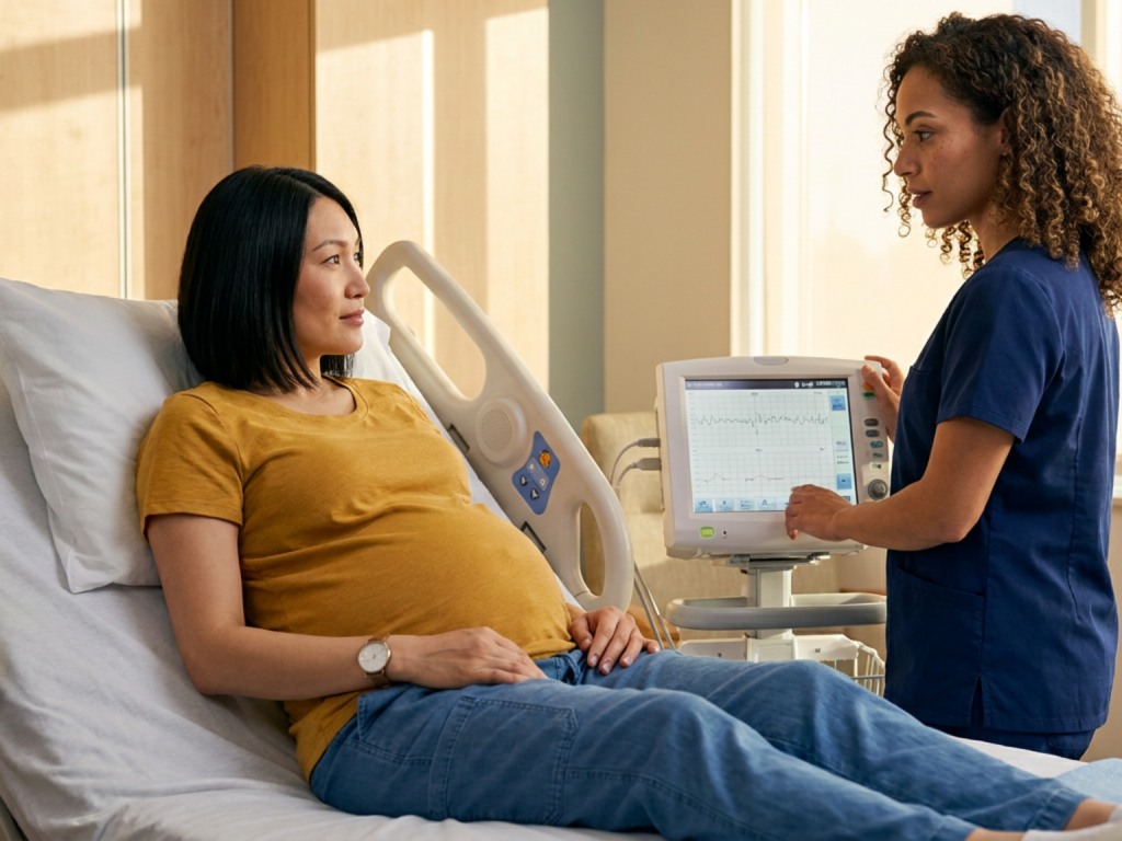
Detection during labor: fetal heart rate monitoring and CTG patterns
During labor, fetal distress is most commonly suspected through fetal heart rate monitoring. Cardiotocography, or CTG, records fetal heart rate and uterine contractions. Monitoring may be intermittent in low-risk labor or continuous when risk factors are present, induction or augmentation is used, epidural-related hypotension occurs, meconium is present, or a concerning pattern has been seen.
Clinicians interpret several features together: baseline fetal heart rate, variability, accelerations, decelerations, and contraction frequency. A normal baseline is generally around 110 to 160 beats per minute. Moderate variability is reassuring because it suggests an intact fetal nervous system and adequate oxygenation. Concerning findings include persistent bradycardia, recurrent late decelerations, recurrent severe variable decelerations, minimal or absent variability, and certain sinusoidal patterns.
Late decelerations are gradual heart rate drops that begin after a contraction starts and recover after it ends. Recurrent late decelerations can suggest uteroplacental insufficiency, especially when variability is reduced. Variable decelerations are often related to cord compression and may be benign if brief with good variability, but recurrent deep or prolonged variables are more concerning. Fetal tachycardia may occur with maternal fever, infection, dehydration, medications, or fetal hypoxia. Interpretation is dynamic; a tracing can improve, worsen, or require urgent action within minutes.
Immediate responses and escalation
When fetal distress is suspected, clinicians often begin intrauterine resuscitation while continuing assessment. Measures may include changing the birthing person’s position to reduce cord or vena cava compression, giving intravenous fluids, treating maternal hypotension, reducing or stopping oxytocin, addressing uterine tachysystole, managing fever, and considering amnioinfusion for recurrent variable decelerations caused by cord compression. Oxygen may be used selectively according to local guidance and the clinical scenario.
If the tracing improves, labor may continue with close observation. If abnormal patterns persist or worsen, the team considers how soon birth can safely occur. In the second stage, an operative vaginal birth decision may be made if the cervix is fully dilated, the fetal head is low enough, and criteria for safety are met. If vaginal birth is not imminent or the situation is severe, cesarean birth indications may include persistent bradycardia, unresolved recurrent late decelerations with reduced variability, suspected abruption, cord prolapse, or other signs of significant fetal compromise.
This escalation can feel sudden for families. A clear explanation helps: what pattern is concerning, what has been tried, what options exist, and why time matters. Even in urgency, patients deserve respectful communication whenever possible.
Risks for the baby and birthing parent
The main fetal risk is inadequate oxygen delivery severe or prolonged enough to cause metabolic acidosis, organ stress, or neurologic injury. Potential outcomes include low Apgar scores, need for newborn resuscitation after birth, admission to neonatal intensive care, hypoxic-ischemic encephalopathy, seizures, meconium aspiration, or, rarely, stillbirth or neonatal death. The actual risk depends on duration, severity, gestational age, and how quickly effective intervention occurs.
For the birthing parent, suspected fetal distress can increase the likelihood of emergency cesarean birth, operative vaginal delivery, anesthesia changes, postpartum hemorrhage, infection risk, and emotional trauma. An emergency birth can be lifesaving and still feel frightening or disappointing. Support after birth, including debriefing with the care team, can help families understand what happened and process the experience.
Not every abnormal fetal heart rate pattern leads to harm. Many babies with non-reassuring tracings are born vigorous, particularly when the pattern is recognized and managed promptly. The purpose of monitoring is to identify the subset of babies who need help before injury occurs. If you are pregnant or in labor and feel worried about fetal movement, bleeding, severe pain, fever, or your baby’s monitoring, ask your team to explain the plan and what would prompt escalation.
Seek urgent medical assessment
- Call maternity triage or emergency services for reduced or absent fetal movements, especially near term.
- Seek immediate care for heavy bleeding, severe abdominal pain, or suspected placental abruption.
- Green or brown amniotic fluid after water breaking should be reported promptly.
- Persistent fever, fainting, severe shortness of breath, or symptoms of shock need urgent evaluation.
- If fetal monitoring is described as abnormal, ask what interventions are being used and what the next step would be.
Tools & Assistance
- Maternity triage phone number or labor and delivery unit
- Kick-count or fetal movement awareness plan recommended by your clinician
- Antenatal testing unit for nonstress tests and ultrasound surveillance
- Birth preference document that includes emergency communication wishes
- Post-birth debrief appointment with the obstetric or midwifery team
FAQ
Is fetal distress the same as a temporary fetal heart rate dip?
No. Brief decelerations can occur in normal labor. Concern rises when patterns are recurrent, prolonged, associated with reduced variability, or occur in a high-risk clinical context.
Can changing position really help?
Sometimes. Position changes may relieve cord compression or improve uterine blood flow. They are usually part of a broader assessment and do not replace urgent delivery if compromise persists.
Does meconium always mean the baby is in danger?
No. Meconium can occur without serious distress, especially near or after term. However, it prompts closer monitoring because it may accompany fetal stress and can affect newborn breathing.
Will fetal distress always mean a cesarean birth?
Not always. Some patterns improve with treatment, and sometimes vaginal birth is close. Cesarean birth may be recommended when concerning patterns persist or birth needs to happen quickly.
What should I ask if my baby’s tracing is non-reassuring?
Ask which feature is concerning, what reversible causes are being treated, whether the tracing is improving, and what criteria would lead to operative or cesarean delivery.
Sources
- Mayo Clinic — Fetal distress: Diagnosis, monitoring, and management
- American College of Obstetricians and Gynecologists — Abnormal fetal heart rate patterns and fetal distress
- National Health Service — Fetal distress: Causes, signs, and treatment
Disclaimer
This article is for educational purposes only and is not a diagnosis or treatment plan. Always seek advice from your obstetrician, midwife, maternity triage unit, or emergency services for pregnancy or labor concerns.