Intro
Week 20 of pregnancy is often described as the halfway point of a 40-week pregnancy. For many people, it is a week filled with mixed emotions: excitement about seeing the baby in detail, anticipation around feeling more consistent movement, and understandable anxiety about the mid-pregnancy anatomy scan.
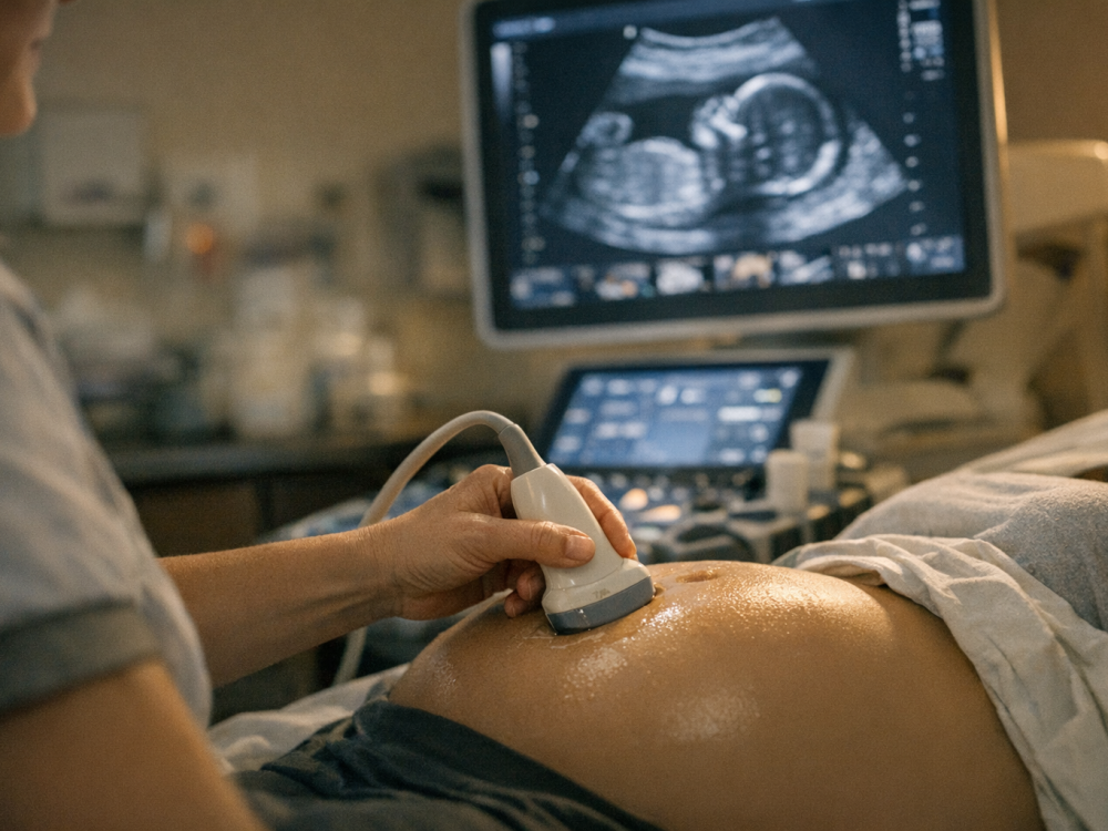
At around 18 to 22 weeks, most pregnancies include a detailed second-trimester ultrasound, often called the 20-week scan, anatomy scan, or anomaly scan. This examination is designed to assess fetal anatomy, growth, placental location, amniotic fluid, and sometimes the cervix and uterine blood flow. It is not a guarantee that every condition will be detected, but it is one of the most clinically important milestones in prenatal care.
Highlights
Week 20 is a symbolic halfway milestone, but fetal development and maternal changes continue at a rapid pace.
The 20-week anatomy scan evaluates major fetal structures such as the brain, heart, spine, kidneys, face, limbs, and abdominal wall.
Fetal movement may be felt as flutters, taps, rolls, or bubbles, but patterns vary depending on placental position, body habitus, and whether this is a first pregnancy.
The scan can be joyful and reassuring, but it can also raise questions or identify findings that need follow-up with a specialist.
Any concerning symptoms, reduced movement later in pregnancy, bleeding, fluid leakage, severe pain, or signs of preeclampsia should be discussed urgently with a healthcare professional.
Why week 20 feels like a turning point
At 20 weeks, pregnancy is approximately halfway through if dating is based on a 40-week gestational age. This midpoint can feel meaningful: the uterus is usually at or near the level of the umbilicus, the pregnancy may be visibly changing the body, and many people begin to feel fetal movement more clearly.
Medically, this stage is important because fetal organs are developed enough to be assessed in more detail by ultrasound, while there is still time to plan additional evaluation, delivery-site decisions, or supportive care if a structural concern is suspected. The second-trimester anatomy survey is typically performed between 18 and 22 weeks because fetal size, organ development, and imaging conditions are often favorable during this window.
Emotionally, the halfway point can bring relief, attachment, and hope, but also vulnerability. It is common to feel nervous before the scan, even if the pregnancy has been uncomplicated. Needing extra images, repeat views, or a referral does not automatically mean something is wrong; fetal position, maternal anatomy, placental location, and technical factors can all affect image quality.
What the 20-week anatomy scan evaluates
The 20-week scan is a detailed ultrasound examination of fetal anatomy. Depending on local protocols and clinical circumstances, the sonographer or clinician may examine the fetal brain and skull, face including the lips, spine, chest, heart, stomach, kidneys, bladder, abdominal wall, limbs, hands and feet, and umbilical cord insertion. Measurements such as head circumference, abdominal circumference, and femur length are often used to assess fetal growth in relation to gestational age.
The fetal heart is a major focus. The scan may assess cardiac position, rhythm, the four-chamber view, and outflow tracts where possible. However, ultrasound cannot detect every congenital heart condition, and some findings are subtle or develop later. If views are incomplete or a concern is identified, a repeat scan or fetal echocardiography may be recommended.
The scan also commonly evaluates the placenta, amniotic fluid, and sometimes the cervix. Placental location matters because a low-lying placenta or placenta previa may require follow-up imaging later in pregnancy. Cervical assessment may be considered if there are risk factors for preterm birth or if local practice includes it. Some centers also assess uterine artery Doppler flow as part of risk evaluation for placental dysfunction, though this varies by setting.
- Brain and skull: ventricles, cerebellum, midline structures, and skull shape may be assessed.
- Spine: alignment and overlying skin contour are examined where visible.
- Heart: chambers, major vessels, rhythm, and position may be reviewed.
- Abdomen: stomach, kidneys, bladder, abdominal wall, and cord insertion are commonly checked.
- Placenta and fluid: placental site and amniotic fluid volume are assessed.
What to expect during the appointment
The appointment may take longer than an early pregnancy scan, often around 30 to 60 minutes, though this varies. The examination is usually transabdominal, using gel and an probe on the abdomen. In some situations, a transvaginal may be used for a more accurate cervical length measurement, placental assessment, or improved visualization.
You may be asked to change position, empty your bladder, or wait while the baby moves into a better position. This can feel frustrating, but it is common. A fetus facing the spine, curled tightly, or moving frequently can make some structures harder to see. A higher body mass index, abdominal scarring, fibroids, or anterior placenta may also affect image clarity.
Some practices allow a support person to attend; others may have restrictions. If you want to know fetal sex and it is visible, you can ask, but the primary purpose of the scan is medical assessment, not sex determination. Conversely, if you do not want to know, tell the sonographer before the examination begins.
Results may be discussed immediately by the sonographer or reviewed later by a radiologist, obstetrician, or maternal-fetal medicine specialist, depending on the healthcare system. If the scan is incomplete, a repeat appointment may simply be scheduled to obtain views that were not possible the first time.
Fetal movement at 20 weeks: what is typical
Around week 20, many pregnant people begin noticing fetal movement, often called quickening. Early movements can feel like flutters, bubbles, popcorn popping, tiny taps, or gentle rolling. People who have been pregnant before may recognize movement earlier, while first-time parents may not identify it until 20 to 24 weeks.
An anterior placenta, meaning the placenta is attached to the front wall of the uterus, can cushion movements and make them harder to feel at first. Fetal position, maternal activity, time of day, and individual sensitivity also influence perception. At this stage, movement is often intermittent and not yet expected to follow a predictable daily pattern.
Formal kick counting is usually emphasized later in pregnancy, often after fetal movement becomes more established. Still, it is reasonable to mention any concerns to your healthcare professional, especially if you previously felt regular movement and then notice a significant change. Local guidance differs, and your maternity team can tell you when and how they recommend monitoring movement.
It can be comforting to remember that an ultrasound may show even when you cannot feel it externally. The nervous system, muscles, and joints are developing coordination, and movements become stronger as the fetus grows.
Understanding scan findings and follow-up
Most 20-week scans do not identify a major structural problem. Sometimes, however, the report may mention an incomplete view, a soft marker, a placental finding, growth measurement variation, or a suspected anomaly. These terms can be alarming, so it is important to ask what the finding means in context and what the next step is.
A soft marker is an finding that is not necessarily a structural abnormality but may slightly change the statistical risk of certain chromosomal conditions when considered alongside age, screening results, and other findings. Examples and interpretation vary, and many isolated soft markers do not change management. Your clinician can integrate ultrasound findings with prior screening such as combined screening, cell-free DNA testing, or diagnostic testing if applicable.
If a structural abnormality is suspected, referral to maternal-fetal medicine, fetal medicine, pediatric cardiology, genetics, or another specialist may be recommended. Further evaluation might include repeat targeted ultrasound, fetal echocardiography, genetic counseling, or diagnostic testing such as amniocentesis. These decisions are individualized and should be made with qualified professionals who can explain benefits, limitations, and risks.
It is also possible to receive uncertain results. Waiting for repeat imaging or specialist review can be emotionally difficult. Bringing a support person, writing down questions, and asking for results in plain language can help you regain a sense of orientation.
Your body at the halfway point
By week 20, common physical experiences include a growing abdomen, round ligament discomfort, lower back or pelvic pressure, heartburn, constipation, nasal congestion, leg cramps, changes in skin pigmentation, and increased vaginal discharge. Many of these symptoms are related to uterine growth, hormonal changes, vascular expansion, and mechanical pressure.
Energy may improve compared with the first trimester, but not always. Sleep can become more disrupted, and emotional responses may be intensified by scan-related anticipation. A balanced approach to self-care can include gentle movement if approved by your clinician, hydration, fiber-rich foods, supportive footwear, rest breaks, and attention to mental health.
At prenatal visits around this time, clinicians may review blood pressure, urine testing if indicated, weight trends, fetal heartbeat, symptoms, blood tests, immunization planning, and any risk factors that need follow-up. If you have a high-risk pregnancy, pre-existing medical condition, prior pregnancy complication, multiple pregnancy, or abnormal screening result, your monitoring schedule may differ.
Questions to ask at the 20-week visit
It is easy to forget questions during a scan or appointment, especially if you feel overwhelmed. Preparing a short list beforehand can help. Consider asking:
- Were all fetal anatomy views completed, or do I need a repeat scan?
- Is fetal growth appropriate for my gestational age?
- Where is the placenta located, and does it require follow-up?
- Is the amniotic fluid volume within the expected range?
- Was the cervix assessed, and is there any concern about cervical length?
- When should I expect fetal movement to become more regular?
- Who will contact me with final results, and when?
- If a finding was seen, what is the most likely explanation and what are the next steps?
You deserve clear, compassionate explanations. If something is unclear, it is appropriate to ask the clinician to repeat it, draw a diagram, provide written information, or explain how urgent the finding is.
When to seek medical advice urgently
- Vaginal bleeding, fluid leakage, or severe abdominal or pelvic pain should be assessed promptly.
- Severe headache, visual symptoms, sudden swelling, chest pain, or shortness of breath may need urgent evaluation.
- Fever, painful urination, or persistent vomiting warrants medical advice.
- If you are concerned about fetal movement, contact your maternity unit or healthcare provider for guidance.
- Do not wait for a routine appointment if symptoms feel severe, sudden, or unusual for you.
Tools & Assistance
- Bring your pregnancy notes, prior screening results, and medication list to the scan appointment.
- Write down questions before the anatomy scan and ask how results will be communicated.
- Use your maternity unit, obstetric clinic, midwife, or maternal-fetal medicine service for individualized advice.
- Consider a support person for emotional backup if your clinic policy allows.
- Track concerning symptoms and timing, but avoid relying on apps or home devices instead of professional assessment.
FAQ
Is the 20-week scan mandatory?
No. It is usually offered as part of routine prenatal care, but you can choose whether to have it. Your clinician can explain what it can and cannot detect.
Can the anatomy scan detect every birth defect?
No. The scan can identify many major structural conditions, but detection depends on the condition, fetal position, image quality, gestational age, and equipment. Some conditions are not visible before birth.
Should I be worried if I do not feel strong movement at 20 weeks?
Not necessarily. Movement perception varies, especially in a first pregnancy or with an anterior placenta. Ask your healthcare provider what is expected for your situation and when to report changes.
What happens if the placenta is low at 20 weeks?
A low-lying placenta often moves away from the cervix as the uterus grows. Follow-up ultrasound later in pregnancy may be recommended to reassess its position.
Can I find out the baby’s sex at the 20-week scan?
Often, yes, if the fetus is positioned clearly and the clinic provides this information. However, the scan’s main purpose is medical evaluation of fetal anatomy and pregnancy structures.
Sources
- NHS — 20-week Screening Scan
- Nebraska Medicine — What to expect during your 20-week prenatal anatomy scan
- PubMed Central — A pictorial guide for the second trimester ultrasound
Disclaimer
This article is for general educational purposes only and is not a substitute for personalized medical care. Always consult your obstetrician, midwife, or qualified healthcare professional about pregnancy symptoms, scan results, or treatment decisions.


Please log in to leave a comment.