
Intro
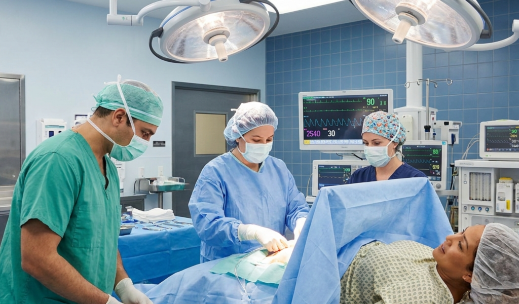
During a cesarean birth, delivering the baby is followed by another essential stage: separating and removing the placenta from the uterus. This usually happens quickly while the surgical team checks the uterus, controls bleeding, and prepares to close the uterine incision.
Most placentas are delivered through the uterine incision by allowing separation to occur naturally and using gentle traction on the umbilical cord. In some circumstances, the surgeon removes the placenta manually. The method depends on the clinical situation, the condition of the mother and baby, the degree of bleeding, and whether the placenta separates as expected.
Highlights
The placenta is removed after the baby is born and before the uterine incision is closed.
Spontaneous separation followed by controlled cord traction is generally preferred when the situation allows.
Manual removal may be necessary when the placenta does not separate, bleeding is significant, or urgent uterine assessment is required.
The team checks that the placenta and membranes appear complete and monitors uterine contraction and blood loss throughout the procedure.
When the placenta is removed
After the baby is delivered through the uterine incision, the umbilical cord is clamped and cut according to the circumstances of the birth. Attention then turns to the placenta, which remains attached to the inner surface of the uterus. The placenta must be removed before the uterus can be repaired and the operation completed.
Placental separation is usually caused by contraction of the uterus. As the uterine muscle contracts, the implantation site becomes smaller and the placenta begins to detach from the uterine wall. The surgeon may observe signs of separation, such as the placenta moving away from the uterine wall or blood appearing behind it. At cesarean delivery, the process may occur without the same visible external signs seen during a vaginal birth because the surgeon has direct access through the uterine incision.
Once the placenta has separated, it is guided out through the incision. The obstetrician then examines the uterine cavity and the placenta before beginning or completing repair of the uterus.
Spontaneous delivery and controlled cord traction
When the mother and baby are stable and there is no urgent reason to remove the placenta immediately, clinicians commonly allow the placenta to separate spontaneously. The surgeon or an assistant may apply gentle, controlled traction to the umbilical cord while supporting the uterus. This technique is often called controlled cord traction.
Controlled cord traction is different from pulling firmly or continuously on the cord. Traction is applied only when separation is likely and is coordinated with uterine contraction. Excessive force could contribute to tissue injury or cord avulsion, so the maneuver is performed by trained clinicians who can respond to resistance.
Evidence summarized in clinical reviews suggests that allowing spontaneous separation with controlled cord traction can reduce blood loss, postoperative hematocrit decline, endometritis, and hospital stay compared with routine manual removal. For this reason, spontaneous placental delivery is generally preferred when it is clinically feasible.
This approach does not mean that the placenta is left unattended. The surgical team continues to assess bleeding, uterine tone, placental completeness, and the mother’s vital signs. If the placenta does not separate promptly or the clinical picture changes, the team may move to another method.
Manual removal of the placenta
Manual removal involves the surgeon placing a gloved hand through the uterine incision, locating the plane between the placenta and the uterine wall, and gently separating the placenta from the implantation site. The placenta is then removed through the incision. This is performed under the anesthesia already being used for the cesarean, so the patient should not experience the procedure in the same way as an awake bedside examination, although sensations of pressure or manipulation may still be possible depending on the anesthetic technique.
Manual removal may be required when the placenta remains attached despite attempts to allow separation, when heavy bleeding suggests that prompt removal is necessary, or when the surgeon needs to inspect the uterine cavity directly. It can also be used when fragments or membranes appear to remain inside the uterus.
Manual removal is not automatically safer simply because it is faster in a particular moment. A meta-analysis of randomized trials found that manual removal was associated with greater blood loss and a higher incidence of endometritis than spontaneous delivery, without a clear reduction in operative time or transfusion requirements. The findings support using manual removal selectively rather than as a routine step for every cesarean.
After removal, the surgeon evaluates the uterine cavity and may use gentle exploration or other measures if retained tissue is suspected. The exact approach depends on bleeding, placental appearance, uterine anatomy, and the suspected cause of incomplete separation.
Checking the placenta and membranes
Once the placenta has been delivered, the team examines its maternal and fetal surfaces. They look for missing cotyledons, torn membranes, abnormal areas, or other evidence that a portion may have remained inside the uterus. This examination is important because retained placental tissue can interfere with uterine contraction and contribute to ongoing bleeding or later infection.
The surgeon also assesses the uterus directly through the incision. The uterine muscle should contract and become firm, a state sometimes described as good uterine tone. A soft or poorly contracted uterus can cause uterine atony, one of the important causes of postpartum hemorrhage. The clinical team responds based on the degree of bleeding and the mother’s condition.
There is no single visual inspection that can guarantee that microscopic placental tissue is absent. However, a careful examination of the placenta, membranes, and uterine cavity helps identify the most obvious problems while the surgical team is still able to address them.
How bleeding is controlled after placental delivery
After the placenta is removed, the uterus normally contracts around the open blood vessels at the implantation site. This contraction acts as a physiological mechanism for reducing blood loss. The team may massage the uterus through the abdominal wall or directly assess it through the incision, depending on the stage of the operation and local practice.
Medication that promotes uterine contraction, often referred to as a uterotonic medication after delivery, is commonly administered as part of routine cesarean care. The choice and dose are individualized because some medicines may be unsuitable for people with particular cardiovascular, hypertensive, respiratory, or other medical conditions. The anesthesiology and obstetric teams coordinate medication, fluid management, blood testing, and monitoring.
If bleeding continues, clinicians look for the cause. Possibilities include uterine atony, trauma to the uterus or surrounding tissues, abnormal placental attachment, retained tissue, or a clotting problem. Management may involve additional uterotonic treatment, surgical repair, removal of retained tissue, uterine compression techniques, blood products, or other procedures. The response is tailored to the situation and follows the hospital’s obstetric hemorrhage protocols.
Blood loss is assessed throughout the operation rather than only at the end. Heart rate, blood pressure, oxygenation, urine output, clinical appearance, and laboratory results can all help the team judge the mother’s status.
When separation is difficult
Sometimes the placenta does not separate along a normal plane. This may occur with abnormal placental attachment, including placenta accreta spectrum, in which placental tissue attaches too deeply into the uterine wall. The risk is higher when the placenta overlies a previous uterine scar or in other specific obstetric circumstances, but risk assessment is individualized.
When abnormal attachment is suspected, forceful attempts to peel the placenta away can cause severe hemorrhage. The surgical plan may therefore differ substantially from routine placental delivery. Depending on prenatal findings, bleeding, fertility goals, available expertise, and emergency conditions, the team may leave the placenta in place, perform additional surgery, or use a planned multidisciplinary approach. These decisions require specialist judgment.
A placenta that is difficult to remove does not necessarily mean that something went wrong. It may reflect the way the placenta is attached or the urgency of the birth. The obstetrician should explain the findings and any additional treatment recommended after the operation, including monitoring for bleeding, infection, or retained tissue.
What the patient may notice afterward
Most patients do not see the placenta being removed because a surgical drape separates the operative field from the upper body. During a cesarean performed with regional anesthesia, such as spinal or epidural anesthesia, a patient may feel pressure, pulling, or movement but should not feel sharp surgical pain. The anesthesiologist can adjust care if discomfort, nausea, anxiety, or other symptoms occur.
After surgery, uterine cramping, vaginal bleeding, fatigue, and soreness are common as the uterus contracts and the body recovers. The amount of bleeding should gradually lessen, but recovery varies. The care team checks the incision, uterine firmness, vital signs, and laboratory results when indicated.
Urgent medical assessment is appropriate for heavy or rapidly increasing bleeding, faintness, shortness of breath, chest pain, severe or worsening abdominal pain, fever, foul-smelling discharge, confusion, or a wound that is opening or producing concerning drainage. These findings can have several causes and should not be self-diagnosed. Contact the maternity unit, obstetric clinician, or emergency service according to the instructions provided at discharge.
When to seek urgent care
- Heavy vaginal bleeding, passing large clots, or bleeding that suddenly increases
- Fainting, marked weakness, rapid heartbeat, shortness of breath, or chest pain
- Fever, chills, worsening abdominal pain, or foul-smelling vaginal discharge
- Increasing redness, swelling, separation, or drainage from the cesarean incision
- Any concern about retained placental tissue or bleeding after discharge
Tools & Assistance
- Ask the obstetric team which placental delivery method was used and whether the placenta appeared complete.
- Request an explanation of any additional procedures, medications, or blood tests performed during the cesarean.
- Keep the maternity unit and emergency contact instructions available after discharge.
- Attend the recommended postpartum follow-up, especially after significant bleeding or difficult placental separation.
FAQ
Is the placenta always removed manually during a cesarean?
No. When circumstances allow, clinicians generally prefer spontaneous separation with gentle controlled cord traction. Manual removal is reserved for situations such as delayed separation, significant bleeding, or suspected retained tissue.
Can I feel the placenta being removed?
With regional anesthesia, you may feel pressure, pulling, or movement, but sharp pain should be treated promptly. Tell the anesthesiologist if you feel pain, nausea, or distress during the operation.
Why does the surgeon inspect the placenta?
The placenta and membranes are checked for missing pieces or abnormalities. This helps the team assess whether tissue may remain in the uterus and whether further evaluation is needed.
Does manual removal increase the risk of infection?
Evidence from randomized trials and meta-analysis associates routine manual removal with a higher incidence of endometritis than spontaneous delivery. Individual circumstances may still make manual removal medically necessary.
What happens if the placenta is abnormally attached?
The team avoids forceful separation when abnormal attachment is suspected because it can cause severe bleeding. Management is individualized and may require specialist surgery, blood-management planning, and close postpartum monitoring.
Sources
- PubMed — Methods of delivering the placenta at caesarean section
- PubMed Central — Manual removal versus spontaneous delivery of the placenta at Cesarean Section: A meta-analysis of randomized controlled trials
- NCBI Bookshelf — Cesarean Delivery
Disclaimer
This article is for general educational purposes and does not replace advice, diagnosis, or treatment from a qualified obstetric or other healthcare professional. Seek urgent medical care for concerning bleeding, pain, fever, breathing difficulty, or other severe symptoms.
















Please log in to leave a comment.